An ear hematoma in cats is a condition characterized by a pocket of blood that forms between the cartilage and skin of the ear flap, also known as the pinna. Imagine the structure of a cat’s ear: a rigid yet flexible cartilage framework that gives the ear its shape, covered by skin on both sides. Within this structure are numerous blood vessels. When these vessels are damaged, blood can leak and accumulate, leading to a noticeable swelling. Because of the cartilage support, cat ears typically stand erect, but a hematoma can distort this natural posture.
Head shaking and scratching are common behaviors in cats, but when excessive, they can become problematic. These actions can cause small separations between the skin and cartilage within the ear. This trauma can also damage the delicate blood vessels in the area. The result is blood seeping into the space created between the cartilage and skin, leading to the formation of a soft, puffy, fluid-filled swelling. This swelling, the hematoma, can vary in size, ranging from a small localized bump to a large mass encompassing the entire ear flap.
Even after a Cat Ear Hematoma heals, there can be lasting changes. Scar tissue and fibrosis, a thickening and scarring of connective tissue, may develop. This can result in a permanent wrinkling or thickening of the ear, often referred to as a “cauliflower ear” appearance.
Recognizing the Symptoms of Cat Ear Hematoma
Cat ear hematomas usually manifest on the inner, or underside, surface of the ear flap. These fluid-filled swellings can be quite small, just a few millimeters in diameter, or they can be extensive, involving the entire pinna. Often, only one ear is affected. When you touch a hematoma, it will feel soft, movable, and compressible. It may also be warm to the touch, and sometimes reddish or painful, depending on the severity and underlying cause.
If your cat has an ear hematoma, you might observe several clinical signs, including:
- Persistent Head Shaking: This is often the most noticeable sign as the cat attempts to relieve discomfort or irritation.
- Excessive Scratching at the Ears: Similar to head shaking, scratching is an attempt to alleviate itchiness or pain.
- Head Tilt: The cat may tilt their head to one side, often towards the affected ear, as a way to manage discomfort.
- Ear Pain: The ear may be sensitive to touch, and the cat may flinch or pull away if you try to examine it.
- Ear Discharge or Odor: If the underlying cause is an ear infection, you might notice discharge or an unusual smell coming from the ear.
- Dirty or Inflamed Ear Canals: Look for redness, debris, or signs of inflammation inside the ear canal.
- Swollen, Red, or Ulcerated Pinna: The ear flap itself may appear swollen, red, and in some cases, ulcerated or raw.
 cat hematoma
Pinpointing the Causes of Cat Ear Hematoma
Trauma resulting from vigorous head shaking or scratching is the most frequent trigger for ear hematomas in cats. This trauma is very often secondary to otitis externa, which is inflammation of the external ear canal. Ear infections are a common cause of otitis externa. The discharge in ear infections is typically composed of bacteria and yeast, but allergies can also play a significant role in predisposing cats to these infections.
Beyond ear infections, other factors can lead to the head shaking and scratching that precipitates hematomas:
- Ear Mites: These tiny parasites are particularly common in kittens and outdoor cats and can cause intense itching and irritation.
- Trauma or Injury: Bites or scratches from fights with other animals, or other forms of direct injury to the ear, can damage blood vessels.
- Foreign Bodies, Polyps, or Cancer: These can cause irritation and inflammation in the ear canal, leading to secondary scratching and head shaking.
- Fragile Blood Vessels: In some medical conditions, such as Cushing’s disease, blood vessels may be more fragile and prone to rupture.
Although less common, immune-mediated conditions could also potentially contribute to hematoma formation.
Diagnosing Cat Ear Hematoma in Felines
Veterinarians typically suspect an ear hematoma based on a physical examination. The characteristic soft, fluid-filled swelling on the ear flap, combined with a history of head shaking or scratching reported by the owner, strongly suggests a hematoma. While the physical exam often confirms the presence of fluid, in some instances, a veterinarian might aspirate a small sample of the fluid with a needle to confirm that it is indeed blood.
It’s important to understand that while fluid analysis can definitively confirm an ear hematoma, it’s not always essential for initiating treatment. In certain situations, draining the blood with a needle can even worsen the condition, potentially leading to the hematoma refilling with more fluid.
To identify the underlying cause of the hematoma, a veterinarian may conduct further diagnostic tests:
- Ear Canal Cytology: Microscopic examination of ear discharge helps identify the presence of yeast, bacteria, or ear mites, common culprits in secondary ear infections.
- Comprehensive Otic Examination: A thorough examination of the ear canal, often using an otoscope, is crucial to rule out foreign objects, growths, or polyps. It also allows visualization of the eardrum to check for any rupture.
- Blood Tests: In some cases, blood tests may be recommended to investigate underlying metabolic diseases that could contribute to fragile blood vessels or other predisposing factors.
Treatment Strategies for Cat Ear Hematoma
Small ear hematomas may sometimes resolve on their own, particularly if the underlying condition, such as an ear infection, is promptly and effectively treated. In these cases, addressing the infection, along with administering anti-inflammatory and pain relief medications, might be sufficient to manage the hematoma.
However, it’s important to note that spontaneous resolution of ear hematomas without veterinary intervention is uncommon. More often, even small hematomas tend to grow larger as they become increasingly irritating, prompting the cat to shake and scratch their head more vigorously.
For larger hematomas, some veterinarians may opt to place an open drain, or cannula. This device allows for continuous drainage of blood from the hematoma. Over time, the cartilage and blood vessels will repair themselves, and the drain can be removed. It’s worth noting that this method can be messy, and pet owners should anticipate needing to use an Elizabethan collar (e-collar) to prevent the cat from interfering with the drain and confining the cat to a designated area to manage drainage.
Another non-surgical approach involves draining the blood from the hematoma and then injecting a steroid medication into the space. While the ear may initially refill with blood, the steroid can help to reduce inflammation and potentially prevent long-term recurrence.
Medications for Cat Ear Hematoma Management
In addition to topical treatments for ear infections, some cats may require systemic antibiotics to combat bacterial infections effectively.
Non-steroidal anti-inflammatory drugs (NSAIDs) like Meloxicam or Onsior are frequently used to manage pain and reduce inflammation within the ear canal. However, it’s crucial to be aware that these medications can have potential side effects and are typically intended for short-term use.
In more severe cases of ear hematoma, corticosteroids may be considered to aggressively decrease inflammation. However, steroids have well-documented side effects in cats, including an increased risk of diabetes, so their use is generally reserved for situations where other treatments are insufficient, and they should be used cautiously.
Cold laser therapy is another treatment modality that may be used to promote healing and reduce inflammation in the affected ear.
Surgical Intervention for Cat Ear Hematoma
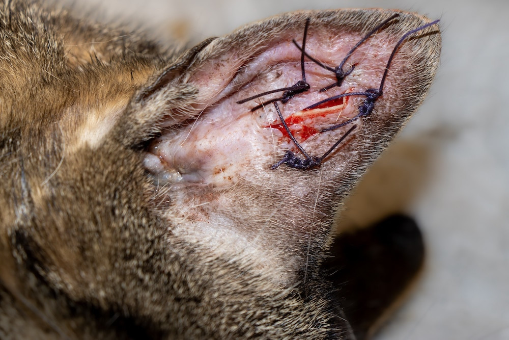
Surgery may be recommended in certain cases, particularly for large or chronic hematomas. A common surgical technique involves making an incision and placing multiple sutures to “tack down” the skin to the cartilage throughout the ear. This helps to eliminate the space where blood can re-accumulate and promotes healing.
Understanding the Cost of Cat Hematoma Surgery
The cost of ear hematoma surgery in cats can vary. If performed by a general practice veterinarian, it may be less than $1,000. However, if a veterinary specialist performs the surgery, the cost could be two to three times higher, depending on the geographic location and the complexity of the case.
Recovery and Ongoing Management After Cat Ear Hematoma Treatment
If an ear infection was present, follow-up appointments to re-evaluate the infection are usually necessary every 1 to 2 weeks until it is fully resolved. Hematomas that are not drained may take longer to be reabsorbed by the body and disappear completely. As long as the underlying cause of the head shaking and scratching is addressed, the hematoma should not worsen. However, complete healing can take several weeks. Recurrence is possible, especially if the underlying cause is not effectively managed.
For cats that undergo surgery, follow-up recheck appointments are needed to monitor healing, along with incision care, typically for around two weeks. It’s also crucial to continue treating any underlying ear infections or other contributing factors during the recovery period.
References
- Tilley LP, Smith FWK. The 5-Minute Veterinary Consult: Canine and Feline. Lippincott Williams & Wilkins; 2005.
- Morgan DVM, DACVIM, DACVO, Rhea V. Veterinary Information Network. Aural Hematoma (Feline). March 2019.
Featured Image: iStock.com/simonkr
References
WRITTEN BY
Lauren Jones, VMDVeterinarian
Dr. Lauren Jones graduated from the University of Pennsylvania School of Veterinary Medicine in 2010, after receiving her bachelor’s degree…